Patient Stories
Walter Suita’s Patient Journey at Tufts Medical Center
April 7, 2020
Walter Suita’s most recent 5K race wasn’t his fastest, but it was one of his biggest victories. Just months earlier, a crushing 13-foot fall had shattered his hip.
Patient Stories
Bob's Story
January 1, 2019
Tufts Medical Center's Weight and Wellness patient describes his journey to stop his diabetes and lose weight.
Patient Stories
Deb Leone's Story
February 1, 2019
Deb was in love with sugar. She decided to cleanse her body of sugar with the help of the Weight and Wellness Center at Tufts Medical Center. Read her story.
Patient Stories
Jabarah Harley's Story
March 1, 2019
Jabarah Harley always struggled with weight management but when the number on the scale changed, she sought out the help of the Tufts Medical Center Weight and Wellness Center team.
Patient Stories
Julia Westover's Story
April 2, 2019
Julia's time as an athlete taught her to never give up and that she didn't like to lose. Julia put those feelings to action when she came to the Weight and Wellness Center at Tufts Medical Center for weight loss surgery.
Articles
Positive Childhood Experiences May Factor in Adult Mental Health
October 1, 2019
Groundbreaking research by Dr. Robert Sege at Tufts Medical Center, indicates that positive childhood experiences may not only decrease the risk of depression or other mental health issues later in life, but may also counteract any detrimental mental health effects of negative or traumatic childhood experiences.
By gtorres, 21 August, 2023

The Department of Dermatology places a big emphasis on redefining what's possible in personalized care for the body's biggest organ — the skin. And just as our skin changes, so do we. Our passion for dermatology leads with layers of comfort, expertise and the curiosity to explore what's next. Between our established experts and up-and-coming dermatologists, we practice a vast scope of specialty services, including:

It's important to note that your health insurance plan will cover a portion of the bill, but there may be additional costs such as deductibles, copayments, coinsurance or services that are not covered. Your policy is a contract between you and your health insurance provider, and it's ultimately your responsibility to pay your hospital bill. 

We offer multiple payment options to assist you in paying your bill. Call the number on your bill to speak with someone about your options.

And if you ever need any assistance with financial arrangements, insurance plans or applying for benefits, our financial counselors are always here to help.

Our facilities offer medically necessary care for all patients round the clock, every day of the year. As frontline caregivers, we provide vital medical care to all individuals who seek our help, regardless of their ability to pay.
By gtorres, 18 August, 2023

When we’re part of a health system that’s nearly as old as the United States itself, our Department of Surgery is bound to make some history along the way. Like, in 1964, when we performed the first-ever small bowel transplant on a person. Or, when researchers Robert Schwartz and William Dameshek discovered chemical 6-mercaptopurine and its immunosuppressive effects, coining the term "immunosuppression” in the 1950s.

Protocol for bone marrow transplantation of mice

Donors: Usually young males, 6-8 weeks of age. Prime with 5-fluorouracil (160-200 mg/kg ) by i.p.. injection. The solution is made by dissolving 100 mg of 5-FU in 400µl of 1N NaOH and then add PBS to 5ml. Harvest bone marrow cells from the femur from day four to day six post-injection.

Harvest: The cells are collected in DMEM with 20 % serum, washed twice with PBS-penicillin streptomycin and resuspended in HBSS for injection.

Recipients: Age 6-10 weeks. Transfer to sterile cages with sterile chow and acidified water (pH 1.3-2.0) 1-2 weeks prior to transplant. Irradiate 8-9 Gy 4 hrs prior to BMT and then again just before transplant. Cells collected just prior to BMT are injected via the tail vein using a 27 G needle. Marrow collected from one mouse can be used to reconstitute the marrow of up to two mice. Recipients are housed in sterile cages post-transplant for an additional two weeks. After that, they can be moved to regular cages. A mortality rate of 10-15% can be expected as a result due to radiation necrosis towards the end of the first-week post-transplantation.


Protocol for subcellular fractionation


  1. Take out half the pancreas and put in 2.2 ml of homogenization buffer (250mM Sucrose, 5mM MOPS, 1mM MgSO4 , pH 6.5 and keep on ice) Note: measure the pH when the buffer is at 4º celsius.
  2. Homogenize the pancreas in the Teflon-coated homogenizer, set at 40 and in reverse. Go slowly up and down 5 times. Transfer the homogenate into 2 eppendorf tubes with 1.1ml in each one (to make duplicates)
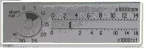
  3. Spin the homogenate for 10 minutes at 150g at 4º Celsius to separate the unbroken cells and other debris from the supernatant. The centrifuge should be set at the lowest speed possible (all the way to the left), you’ll only see one marker. When it has started, turn the knob one ’click’ until there are two markers visible in the display. 
Subcellular 1
  1. Meanwhile, for each sample, put 500µL of 200mM sodium acetate buffer(mW=136.1: 1.361g in 50mL deionized water, pH5.0 at 37º C) together with 400µL 6.25mM p-Nitrocatechol Sulfate Solution (mW=311.36: 19.4g in 10mL deionized water)(PNCS) in a 12 well plate at 37º C in a shakingbath (100 rpm). Leave it long enough to completely warm to 37º C until further process.
  2. After spinning, take 750µL from the supernatant (and not all), this way, the chances that you’ll take some of the pellet are minimal. Be careful not to take any debris out. This 750µL is considered the total. Discard the pellet.
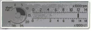
  3. Spin the supernatant at 1000g for 10 minutes at 4º Celsius (set the speed exactly as indicated in the picture). 
Subcellular 2
  1. Separate the supernatant from the pellet very carefully and try to take the whole and only the supernatant, this requires precise pipetting. The pellet is considered the zymogen enriched fraction (ZRF). Put this on ice again
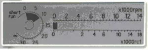
  2. Respin the supernatant for 15 minutes at 12.000g at 4º Celsius (see image). The pellet remaining after the spin is the Lysosomal enriched fraction (LRF). If you only going to read the ZRF and the LRF you can discard the supernatant from this tube. 
Subcellular 3
  1. After putting the tubes in the centrifuge for the LRF spin, proceed with the preparation for the actual assay for aryl sulfatase; To the ZRF pellet add 150 µL of ice-cold sodium chloride solution (0.2% w/v in deionized water). Resuspend the pellet in this buffer very thoroughly by pipetting up and down (p1000) and make sure you don’t see any packed particles anymore.
  2. From the resuspended (enzyme) solution, take 100 µL and add this to the prewarmed acetate/PNCS buffer and set the timer to 30 minutes.
  3. Prepare cups for each well containing 5mL 1M sodium hydroxide (NaOH). This is used to stop the reaction.
  4. After exactly 30 minutes, transfer the buffer containing the enzyme to the NaOH cups, this will stop the reaction. From this 6mL take 1mL and read it in the spectrophotometer with ? set at 515nm. Before using the spectrophotometer you have to blank it. To prepare the blank; take a warm (37º C) mixture of sodium acetate and PNCS, add this to 5mL of 1M NaOH. After this, add 100 µL of any sample (or combined sample) and mix gently. From this solution, take 1mL, this is your blank.
  5. repeat step 9-12 for the LRF.

Note: process the fractions as soon as possible, although aryl sulfatase is considered very stable, I’ve noticed a decrease if left unprocessed for a while. So don’t wait until you’ve span the LRF,

Display title
Methods
Jump back to top